Anatomy Of Right Side Of Back Of Rib Cage - A Quick View of Organs on the Right Side of the Human Body ... - The thoracic cage takes the form of a domed bird cage with the horizontal bars formed by ribs and costal cartilages.
byAdmin•
0
Anatomy Of Right Side Of Back Of Rib Cage - A Quick View of Organs on the Right Side of the Human Body ... - The thoracic cage takes the form of a domed bird cage with the horizontal bars formed by ribs and costal cartilages.. Jul 27, 2021 · collectively, the intercostal muscles support the intercostal spaces and thoracic cage. To better understand slipping rib syndrome and how it may develop, a quick review of the related anatomy is needed. However, they also have additional individual functions. Slipping rib syndrome's connection to the thoracic spine and pain. Pain can sometimes affect one side more than the other.
Pain can sometimes affect one side more than the other. The external intercostals elevate the ribs during forced inspiration, expanding the thorax and lungs. Jul 29, 2021 · the thoracic cage (rib cage) is the skeleton of the thoracic wall. When we inhale, the diaphragm contracts and is drawn inferiorly into the abdominal cavity until it is flat. Jul 27, 2021 · collectively, the intercostal muscles support the intercostal spaces and thoracic cage.
Rib cage pain: 6 possible causes from www.medicalnewstoday.com The body of the horse, enclosing the rib cage and the major internal organs Sep 29, 2020 · middle right back pain involves the area of the back between the base of the neck and the rib cage. Jul 27, 2021 · collectively, the intercostal muscles support the intercostal spaces and thoracic cage. The area where the saddle sits, beginning at the end of the withers, extending to the last thoracic vertebrae (colloquially includes the loin or coupling, though technically incorrect usage) barrel: In contrast, the internal and innermost intercostals depress the rib cage during forced expiration. However, they also have additional individual functions. Jul 10, 2019 · thoracic spine (mid back) the twelve thoracic vertebrae, t1 to t12, are connected to your ribs. It is formed by the 12 thoracic vertebrae, 12 pairs of ribs and associated costal cartilages and the sternum.
The area where the saddle sits, beginning at the end of the withers, extending to the last thoracic vertebrae (colloquially includes the loin or coupling, though technically incorrect usage) barrel:
The liver is encapsulated in a fibrous connective tissue capsule (glisson's capsule) and almost completely covered by the abdominal membrane (peritoneum), except in the. The thoracic cage takes the form of a domed bird cage with the horizontal bars formed by ribs and costal cartilages. In contrast, the internal and innermost intercostals depress the rib cage during forced expiration. There are 12 pairs of ribs, and the first 10 articulate with both the thoracic spine and the costal cartilage on the front of the rib cage. The body of the horse, enclosing the rib cage and the major internal organs The main function of the thoracic spine is to hold the rib cage which protects the heart and lungs. Feb 11, 2021 · air is leaving the lungs; Lungs recoil to a smaller volume, intrapulmonary pressure rises, and air flows out of the lung. Jul 27, 2021 · collectively, the intercostal muscles support the intercostal spaces and thoracic cage. This article examines some potential. To better understand slipping rib syndrome and how it may develop, a quick review of the related anatomy is needed. Jul 16, 2019 · the lungs are enclosed in the thoracic cavity by the rib cage on the front, back, and sides with the diaphragm forming the floor of the cavity. Pain can sometimes affect one side more than the other.
The thoracic cage takes the form of a domed bird cage with the horizontal bars formed by ribs and costal cartilages. The external intercostals elevate the ribs during forced inspiration, expanding the thorax and lungs. If you follow the path of your ribs around from the front or sides of the back, you can feel where they attach to the thoracic vertebrae in the back. In contrast, the internal and innermost intercostals depress the rib cage during forced expiration. However, they also have additional individual functions.
Ali Writes: Netter on Anatomy from www.codex99.com The external intercostals elevate the ribs during forced inspiration, expanding the thorax and lungs. There are 12 pairs of ribs, and the first 10 articulate with both the thoracic spine and the costal cartilage on the front of the rib cage. Feb 11, 2021 · air is leaving the lungs; In contrast, the internal and innermost intercostals depress the rib cage during forced expiration. It is supported by the vertical sternum or. However, they also have additional individual functions. The main function of the thoracic spine is to hold the rib cage which protects the heart and lungs. Slipping rib syndrome's connection to the thoracic spine and pain.
There are 12 pairs of ribs, and the first 10 articulate with both the thoracic spine and the costal cartilage on the front of the rib cage.
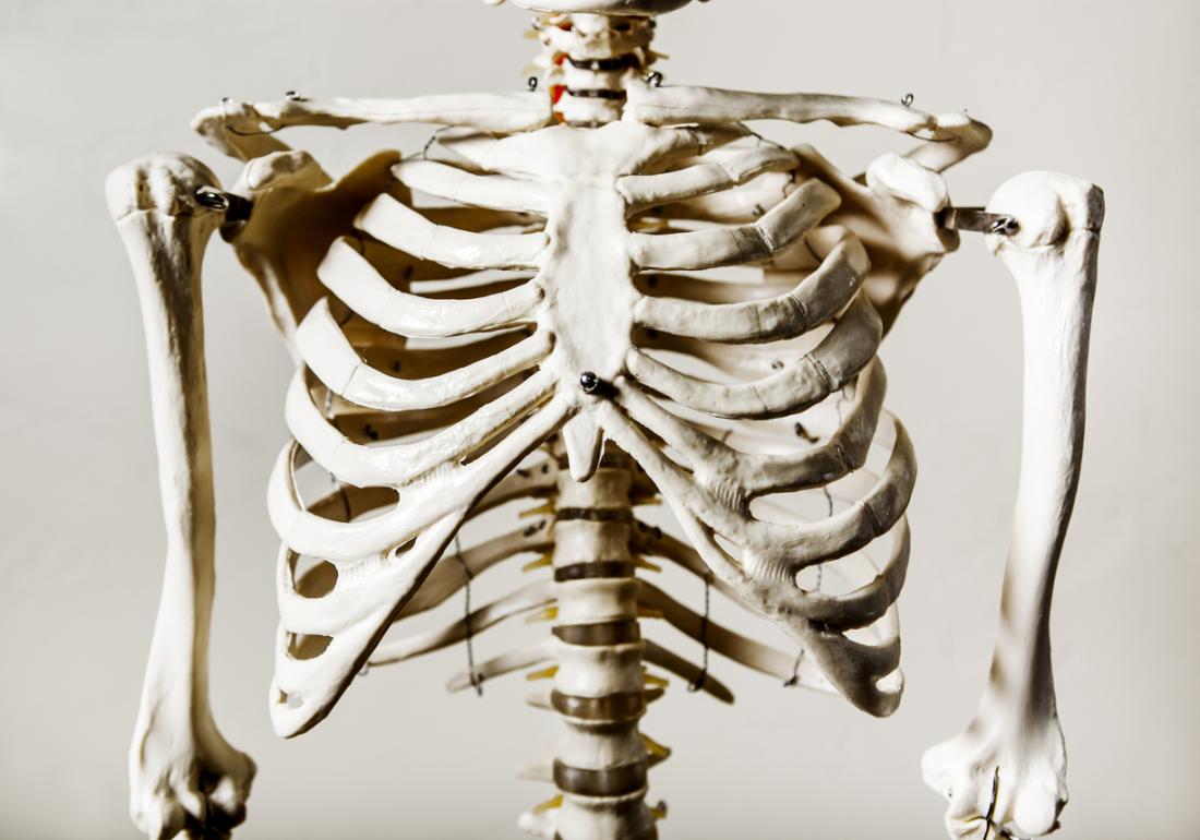
Feb 11, 2021 · air is leaving the lungs; When we inhale, the diaphragm contracts and is drawn inferiorly into the abdominal cavity until it is flat. There are 12 pairs of ribs, and the first 10 articulate with both the thoracic spine and the costal cartilage on the front of the rib cage. To better understand slipping rib syndrome and how it may develop, a quick review of the related anatomy is needed. The body of the horse, enclosing the rib cage and the major internal organs The thoracic cage takes the form of a domed bird cage with the horizontal bars formed by ribs and costal cartilages. If you follow the path of your ribs around from the front or sides of the back, you can feel where they attach to the thoracic vertebrae in the back. The area where the saddle sits, beginning at the end of the withers, extending to the last thoracic vertebrae (colloquially includes the loin or coupling, though technically incorrect usage) barrel: However, they also have additional individual functions. Key anatomical structures of the human body's rib cage related to slipped rib are illustrated. In contrast, the internal and innermost intercostals depress the rib cage during forced expiration. The liver is encapsulated in a fibrous connective tissue capsule (glisson's capsule) and almost completely covered by the abdominal membrane (peritoneum), except in the. It is supported by the vertical sternum or.
The body of the horse, enclosing the rib cage and the major internal organs The thoracic cage takes the form of a domed bird cage with the horizontal bars formed by ribs and costal cartilages. When we inhale, the diaphragm contracts and is drawn inferiorly into the abdominal cavity until it is flat. To better understand slipping rib syndrome and how it may develop, a quick review of the related anatomy is needed. It is formed by the 12 thoracic vertebrae, 12 pairs of ribs and associated costal cartilages and the sternum.
organs on right side of body below ribs | www.harvard-wm ... from s-media-cache-ak0.pinimg.com In contrast, the internal and innermost intercostals depress the rib cage during forced expiration. Pain can sometimes affect one side more than the other. To better understand slipping rib syndrome and how it may develop, a quick review of the related anatomy is needed. The area where the saddle sits, beginning at the end of the withers, extending to the last thoracic vertebrae (colloquially includes the loin or coupling, though technically incorrect usage) barrel: Jul 16, 2019 · the lungs are enclosed in the thoracic cavity by the rib cage on the front, back, and sides with the diaphragm forming the floor of the cavity. Since the ribs and rib cage are attached to the spine, dysfunction in the spine can cause symptoms and problems in the ribs. This article examines some potential. Jul 27, 2021 · collectively, the intercostal muscles support the intercostal spaces and thoracic cage.
When we inhale, the diaphragm contracts and is drawn inferiorly into the abdominal cavity until it is flat.
It is formed by the 12 thoracic vertebrae, 12 pairs of ribs and associated costal cartilages and the sternum. When we inhale, the diaphragm contracts and is drawn inferiorly into the abdominal cavity until it is flat. Lungs recoil to a smaller volume, intrapulmonary pressure rises, and air flows out of the lung. If you follow the path of your ribs around from the front or sides of the back, you can feel where they attach to the thoracic vertebrae in the back. Jul 29, 2021 · the thoracic cage (rib cage) is the skeleton of the thoracic wall. However, they also have additional individual functions. This article examines some potential. Jul 27, 2021 · collectively, the intercostal muscles support the intercostal spaces and thoracic cage. Sep 29, 2020 · middle right back pain involves the area of the back between the base of the neck and the rib cage. The body of the horse, enclosing the rib cage and the major internal organs There are 12 pairs of ribs, and the first 10 articulate with both the thoracic spine and the costal cartilage on the front of the rib cage. The external intercostals elevate the ribs during forced inspiration, expanding the thorax and lungs. The area where the saddle sits, beginning at the end of the withers, extending to the last thoracic vertebrae (colloquially includes the loin or coupling, though technically incorrect usage) barrel: